Cellular elements in the CNS glial cells
For many years following their discovery, glial cells were viewed as CNS connective tissue. The word glia is Greek for glue. However, today these cells are recognized for their role in communication within the CNS in partnership with neurons. Unlike neurons, glial cells continue to undergo cell division in adulthood, and their ability to increase is particularly noticeable after brain injury.
Neurons
Neurons in the mammalian central nervous system come in many different shapes and sizes. The cell body (soma) contains the nucleus and is the metabolic center of the neuron. Neurons have several dendrites that extend outward from the cell body and arborize extensively. Particularly in the cerebral and cerebellar cortex, the dendrites have small knobby projections called dendritic spines. A typical neuron also has a long fibrous axon that originates from a somewhat thickened area of the cell body, the axon hillock.
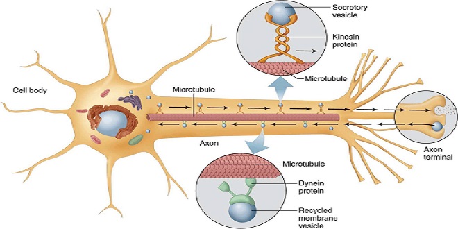
Axonal transport
Neurons are secretory cells, but they differ from other secretory cells in that the secretory zone is generally at the end of the axon, far removed from the cell body. The apparatus for protein synthesis is located mainly in the cell body, with the transport of proteins and polypeptides to the axonal ending by the axoplasmic flow.
Excitation & conduction
Nerve cells have a low threshold for excitation. The stimulus may be electrical, chemical, or mechanical. As a result, two types of physicochemical disturbances are produced: local, no propagated potentials called, depending on their location, synaptic, generator, or electrotonic potentials; and propagated potentials, the action potentials (or nerve impulses).
Ionic fluxes during the action potential
The cell membranes of nerves, like those of other cells, contain many different types of ion channels. Some of these are voltage-gated, and others are ligand-gated. It is the behavior of these channels, particularly Na+ and K+ channels, which explains the electrical events in nerves.
Distribution of ion channels in myelinated neurons
The spatial distribution of ion channels along the axon plays a vital role in initiating and regulating the action potential. Voltage-gated Na+ media are highly concentrated in the nodes of Ranvier and the initial segment in militated neurons. The initial part and, in sensory neurons, the first node of Ranvier, are the sites where impulses are usually generated. The other nodes of Ranvier are the sites to which the impulses jump during salutatory conduction.
“All-or-none” law
It is possible to determine the minimal intensity of stimulating current (threshold intensity) that, acting for a given duration, will produce an action potential. The threshold intensity varies with the time; with weak stimuli, it is long, and with solid inspirations, it is short. The relation between the strength and the duration of a threshold stimulus is called the strength-duration curve.
Electrotonic potentials, local response, & firing level
Although subthreshold stimuli do not produce an action potential, they affect the membrane potential. This can be demonstrated by placing recording electrodes within a few millimeters of a stimulating electrode and applying sub-threshold stimuli of fixed duration.
Changes in excitability during electrotonic potentials & the action potential
During the action potential, as well as during electrotonic potentials and the local response, the neuron’s threshold to stimulation changes. Hyperpolarizing responses elevate the point, and depolarizing potentials lower it as they move the membrane potential closer to the firing level. During the local reaction, the threshold is reduced, but during the rising and much of the falling phases of the spike potential, the neuron is refractory to stimulation.
Summary
There are two main types of microglia and microglia. Microglia are scavenger cells. Microglia include oligodendrocytes, Schwann cells, and astrocytes. The first two are involved in myelin formation; astrocytes produce substances that are tropical to neurons, and they help maintain the appropriate concentration of ions and neurotransmitters.